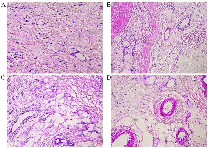
Figure 4.
(A) Histopathological examination of the first choke zone in the surgical flaps showed that the degree of inflammatory cell infiltration and interstitial edema was not obvious in the control group. (B) In the experimental group, tissue clearance showed edema, inflammatory cell aggregation and vessel dilation at 3 days post operation. (C and D) The dilation of choke vessel caliber and increasing vessel wall thickness was clear up to 7 days following flap elevation (C) Results at 5 days (D) results at 7 days. Samples were stained with hematoxylin and eosin. (magnification, ×100).

