Figure 3.
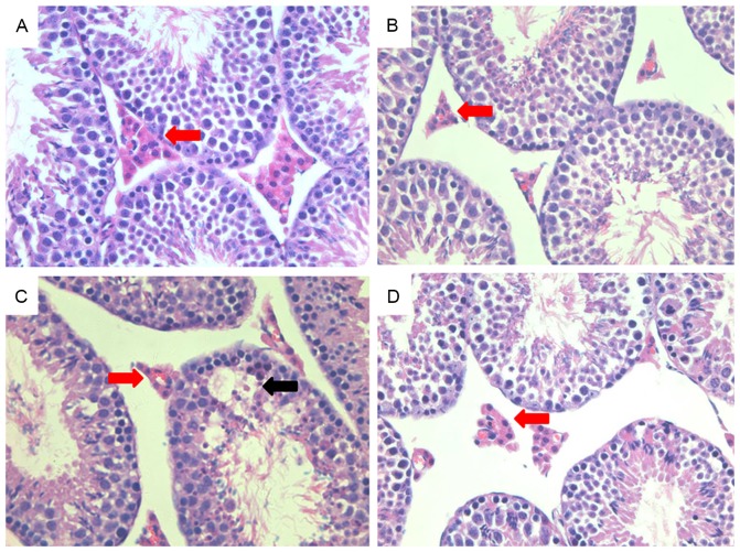
Interstitial morphology of mouse testes. Testicular tissue from each group was paraffin-embedded and stained with hematoxylin and eosin. (A) The intact structure of testicular tissue was clearly evident in the control group, with large numbers of Leydig cells present. (B) Tissue morphology appeared similar to that in the control tissue after treatment roscovitine and subsequently with LPS. (C) In testicular tissue from mice treated with LPS only, few Leydig cells were detected and tissue morphology was altered (black arrow). (D) After treatment with DMSO vehicle only, the structure was similar to that in the control group. Magnification ×200. Leydig cells are indicated by red arrows. LPS, lipopolysaccharide; DMSO, dimethyl sulfoxide.

