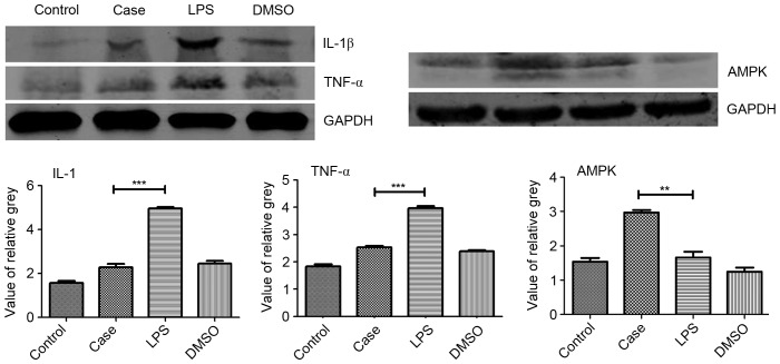
Figure 5.
Protein expression of IL-1β, TNF-α and AMPK. Protein levels of IL-1β, TNF-α and AMPK were determined by western blot analysis of testicular tissue from each group 12 h after each treatment. Roscovitine was found to significantly increase the expression of AMPK compared with that in the other three groups. Furthermore, IL-1β and TNF-α expression in the case group were significantly downregulated compared with that in the LPS group. **P<0.01; ***P<0.001. Groups: Control, injected with saline; case, injected with roscovitine (5 mg/kg) dissolved in DMSO followed by injection of 5 mg/kg LPS in saline; LPS, injected with LPS only; DMSO, injected with DMSO only; LPS, lipopolysaccharide; DMSO, dimethyl sulfoxide; TNF, tumor necrosis factor; IL, interleukin; AMPK, adenosine monophosphate-activated protein kinase.

