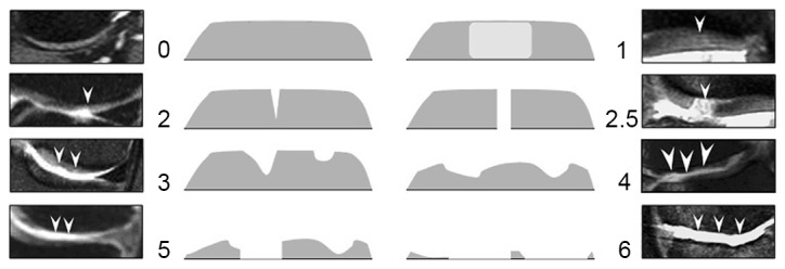
Figure 1.

Cartilage signal intensity and morphology were scored using an 8-point scale from Hayashi et al (13): 0, normal thickness and signal; 1, normal thickness but increased signal on fat-suppression proton density-weighted turbo spin echo image; 2.0, partial-thickness focal defect <1 cm in greatest width; 2.5, full-thickness focal defect <1 cm in greatest width; 3, multiple areas of partial-thickness (grade 2.0) defects intermixed with areas of normal thickness, or a grade 2.0 defect >1 cm comprising <75% of the subregion; 4, diffuse (≥75% of the subregion) partial-thiwness loss; 5, multiple areas of full-thickness loss (grade 2.5), or a grade 2.5 lesion >1 cm comprising <75% of the subregion; 6, diffuse (≥75% of the subregion) full-thickness loss. Arrowheads indicate cartilage damage degree from 0 to 6 standard atlas in cases.
