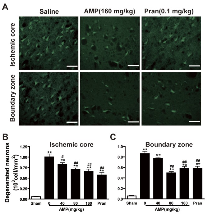
Figure 5.
Effects of AMP and pran on the density of degenerated neurons in cortex 24 h following middle cerebral artery occlusion in rats. Brain sections were stained with Fluoro-Jade B fluorescent dye to detect degenerated neurons 24 h following reperfusion. (A) AMP (160 mg/kg, p.o.) and pran (0.1 mg/kg, i.p.) reduced the density of Fluoro-Jade B-stained neurons in the ischemic core (top panels) and boundary zone (bottom panels). Scale bar, 100 µm. Numbers of Fluoro-Jade B-stained neurons in (B) the ischemic core and (C) boundary zone are summarized. AMP (80 and 160 mg/kg, p.o.) and pran (0.1 mg/kg, i.p.) significantly reduced the number of degenerated neurons of the ischemic core and boundary zones (n=6). **P<0.01 vs. sham; #P<0.05 and ##P<0.01 vs. ischemic control, analyzed by one-way analysis of variance. AMP, ampelopsin; Pran, pranlukast.

