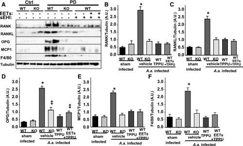
Fig. 3.
The dysregulated RANK/RANKL/OPG system in PD is restored by chemical or genetic ablation of sEH. Protein expression levels of osteoclastogenesis-related factors in gingival tissues from all experimental groups were investigated by Western blotting. For quantification, band intensity was normalized to that of α-tubulin. Protein band intensity is represented as arbitrary units. Density quantification included all animals, and mean ± S.E.M. of each group (n = 6 per group) is displayed in the bar graphs. (A) Original blots displaying two randomly selected animals. (B) Bar graphs of mean band intensity for RANK (B), RANKL (C), OPG (D), MCP-1 (E) measured for all six mice and F4/80 (F) (*P < 0.001, ‡P < 0.03, one-way ANOVA followed by Student’s Newman–Keuls post hoc all pairwise comparison). WT, wild type.

