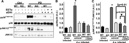
Fig. 4.
PD-mediated phosphorylation of proinflammatory p38 and JNK1/2 is reduced by chemical or genetic ablation of sEH. Phosphorylation and activation of p38 and JNK1/2 were quantified from all groups by normalizing band intensity to that of α-tubulin. (A) Original blots displaying two randomly selected animals. (B and C). Bar graphs of phosphorylation status of p38 and JNK1/2. Mean band intensity is measured for all six mice and is represented as arbitrary units (mean ± S.E.M.) (*P < 0.001, ‡P = 0.01, ▾P = 0.024, one-way ANOVA followed by Student’s Newman–Keuls post hoc all pairwise comparison). WT, wild type.

