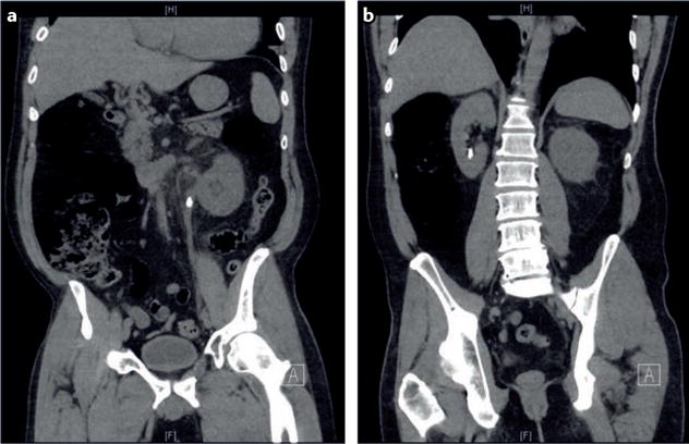
Figure 2. A coronal demonstration of bilateral 8 mm nephrolithiasis on noncontrast CT.

These stones are clearly visible using this imaging modality. Additional anatomical detail can be obtained by reconstructing the images in an axial plane. a | This coronal CT image clearly demonstrates a left-sided obstructing stone. b | Posterior coronal CT view of panel a demonstrating a lower-pole nonobstructing stone. An excellent level of anatomical detail can be seen here and can be further increased by reconstructing the image in an axial plane.
