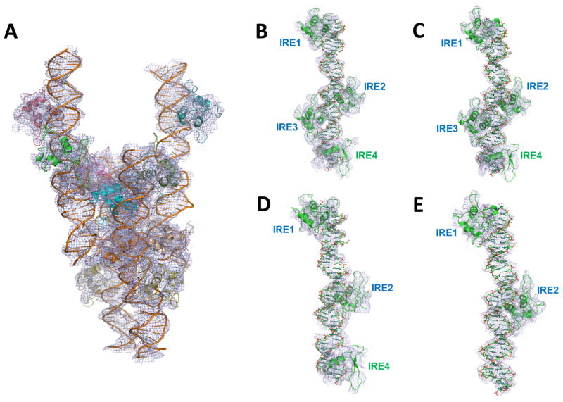
Figure 4. Low resolution crystal structure of the FoxO1-DBD-40mer DNA and the makeup of each complex within the asymmetric unit.
(A) Low resolution (5.0 Ǻ) crystal structure of the 40mer complex. The entire asymmetric unit content with four independent complexes is shown in which each complex contains a different number of FoxO1 proteins bound to DNA containing all four IRE sites. (B–E) Makeup of each complex. Two complexes had all IRE sites occupied (B–C) while two other complexes had partially occupied IRE sites (D: 3 sites and E: 2 sites). Foxo1-bound IRE sites in each complex are labeled. Electron density maps were contoured at 0.8σ.

