
Figure 1.
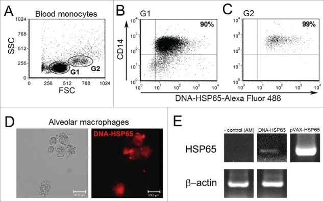
Uptake of DNA-HSP65 by monocytes and alveolar macrophages. Purified CD14+ cells, from six healthy individuals, and alveolar macrophages (AM) were stimulated for 4 hours with Alexa Fluor labeled DNA-HSP65 and analyzed by flow cytometry or fluorescence microscopy, respectively. (A) Cells were gated by forward (FSC) and side (SSC)-scatter, and analysis was performed on gate 1 (G1), small CD14+ monocytes, and gate 2 (G2), large CD14+ monocytes. (B and C) Percentage of double-positive cells (CD14+/DNA-HSP65-Alexa Fluor 488+) for G1 (B) and G2 (C). (D) AM were analyzed by differential interference contrast microscopy and fluorescence microscopy. By RT-PCR (E) Expression of mycobacterial Hsp65 mRNA by unstimulated AM (negative control), DNA-HSP65-stimulated AM, pVAX-HSP65 (positive control).