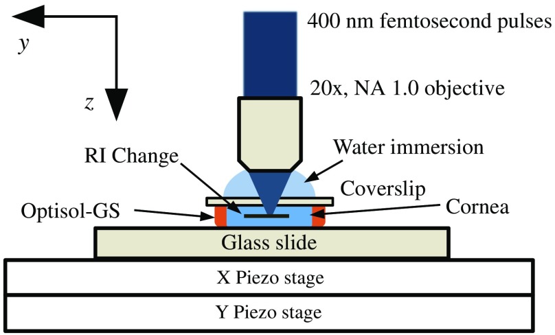
Fig. 1.
Schematic of the blue-IRIS experimental setup. Pulses of 800 nm, 100 fs from a Ti:Sapphire laser were frequency doubled to produce 400 nm, 100 fs pulses using second harmonic generation. These pulses were then tightly focused through a , water immersion objective into the stromal region of an excised corneal sample mounted on a glass slide, bathed in a storage medium (Optisol-GS, Bausch & Lomb), and applanated with a #1 coverslip. The whole assembly was placed on a three-dimensional scanning platform. The piezostages were used to inscribe the IRIS phase grating pattern by raster scanning the sample under the microscope objective. The scan speed (3, 5, for feline tissue and for human tissue) describes the speed of the -axis piezo stage.

