
Figure 1.
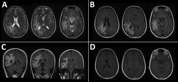
Magnetic resonance imaging of the brain of a 46-year-old immunocompromised woman with central nervous system brucellosis granuloma and white matter disease, Saudi Arabia. A) Axial T2 images showing hyperintensity in the right frontoparietal lobe and right temporal lobe. B) Axial fluid-attenuated inversion recovery (FLAIR) and C) coronal FLAIR images showing that hypersensitivity extends to U-fibers without involvement of the cortex. D) Gadolinium-enhanced image showing that no appreciable mass effect and no central or peripheral enhancement after administration of gadolinium were observed. Each image within each panel shows involvement in different levels of frontal, parietal, and temporal lobes.