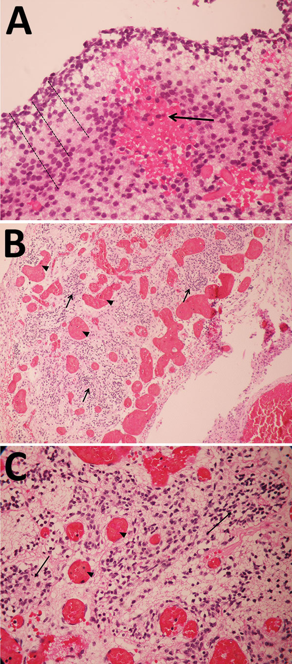
Figure 1.

Pathology findings for case 1, involving a fetus examined after pregnancy termination who had severe neurologic defects attributed to maternal Zika virus infection, Colombia. A) Remnant tissue of cerebral cortex showing a reduced neuroblast layer (dotted lines) and hemorrhagic foci (arrow). Hematoxylin and eosin (H&E) staining; original magnification ×40. B) Glial leptomeningeal heterotopy showing congestive blood vessels (arrowhead) and foci of glial heterotopia (arrows). H&E staining; original magnification ×10. C) Glial leptomeningeal heterotopy showing congestive blood vessels (arrowhead) and foci of glial heterotopia (arrows). H&E staining; original magnification ×40.
