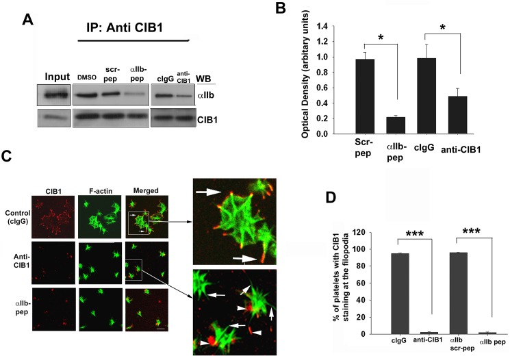
Fig 2. Association of CIB1 with αIIb is needed for platelet spreading but not filopodia formation.
(A) Western blot analysis of anti-CIB1 immunoprecipitates of lysates from platelets incorporated with either αIIb peptide (2.5 μM) or anti-CIB1 antibody (0.1 mg/ml) as indicated. DMSO, scrambled αIIb peptide (2.5 μM) and irrelevant isotype specific antibody (cIgG, 0.1 mg/ml) were used as control. Upper blot shows the co-immunoprecipitation of αIIb. Same blot was reprobed with anti-CIB1 to ensure equal amount of protein in the immunoprecipitates (lower blot). Input represents sample of platelet lysate prior to immunoprecipitation. (B) Quantitation of αIIb bands from (A) normalized to corresponding CIB1 band. (*P<0.05) (C) Confocal images of platelets incorporated with αIIb peptide, anti-CIB1 or corresponding controls DMSO, scrambled αIIb peptide, or cIgG and were allowed to spread on Fg for 45 min and stained for F-actin; boxed area is enlarged to visualize colocalization (arrows); non-specific red speckles are shown by arrowheads; original magnification, X 1600. (D) Quantitation of platelets from C showing CIB1 staining at the filopodia (***P<0.001). Error bars indicate mean ± SEM of at least three independent experiments.

