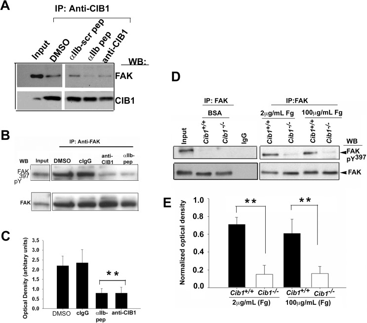
Fig 4. Association of CIB1 with αIIb is essential for the recruitment and activation of FAK.
(A) Western blot analysis of anti-CIB1 immunoprecipitates of lysates of platelets incorporated with anti-CIB1 or αIIb peptide and corresponding scrambled peptide or DMSO and allowed to spread on Fg. Input represents sample of platelet lysates prior to immunoprecipitation. FAK bands are shown in upper blot. Same blot was reprobed with anti-CIB1 to ensure an equal amount of protein in the precipitates (lower blot). (B) Western blot analysis of anti-FAK immunoprecipitates of lysates of platelets treated as above. cIgG was used as control. Activated FAK bands identified using anti-pY397 are shown in upper blot. The same blot was reprobed for total FAK to ensure an equal amount of protein in the precipitates. (C) Quantitation of optical density of phosphorylated FAK bands from B, normalized to corresponding total FAK band (**P<0.01). Error bars indicate mean ± SEM of at least three independent experiments. (D) Western blot analysis of anti-FAK immunoprecipitate of lysates of Cib1+/+ and Cib1-/- mouse platelets exposed to BSA or immobilized Fg. Activated FAK bands identified using anti-pY397 are shown in upper blot. The same blot was reprobed for total FAK to ensure equal protein in the precipitates. (E) Quantitation of optical density of phosphorylated FAK bands from D, normalized to corresponding total FAK band. (**P<0.01).

