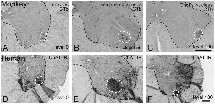
Fig 1. Identification of rostral and caudal landmarks in the lumbosacral enlargement in rhesus monkey and humans.
(A-C) Retrogradely labeled motoneurons in the rhesus monkey lumbosacral cord following CTb injections into the (A) iliopsoas, (B) semimembranosus, and (C) external sphincter muscle. Note that the iliopsoas motoneuron pool demarcates the rostral end of the enlargement, where the ventral horn extends laterally. The caudal landmark is demarcated by pelvic floor motoneurons of the external sphincter (Onuf’s nucleus) touching the edge of the ventral gray matter. (D-F) ChAT-IR neurons in the human lumbosacral cord (87 year old woman with Dementia with Lewy Bodies). Panels (D-F) are homologous to panels (A-C) in the monkey. Note that the changes in shape of the ventral horn, demarcating the rostral and caudal landmarks, can be identified in the human spinal cord. Similar to monkey, these changes in shape are determined by (ChAT-IR) motoneurons, similar to the rhesus monkey. Bars in (A-C), and (D-F) = 500μm.

