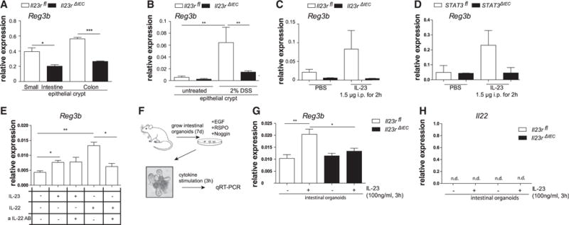
Figure 5. Epithelial IL-23R Orchestrates Reg3b Expression.

(A) Small intestinal and colon crypts were isolated, and gene expression of Reg3b was assessed. Data are representative of at least 3 animals/genotype.
(B) Expression of Reg3b in response to DSS induction was quantified in colonic epithelial cells isolated after 3 days of 2% DSS colitis induction.
(C and D) Il23rfl and Il23rΔIEC (C) and STAT3fl and STAT3ΔIEC (D) animals were injected with IL-23 (1.5 μg) for 2 hr. Colon transcript levels of Reg3b were assessed by real-time PCR.
(E) Freshly isolated epithelial cells from WT animals were stimulated with IL-23 (50 ng/ml) or IL-22 (50 ng/ml) in the presence or absence of neutralizing αIL-22 antibody (1 μg/ml) for 3 hr. Transcript levels of Reg3b were assessed by real-time PCR.
(F) Cultivation of intestinal organoids from Il23rfl and Il23rΔIEC mice. (G and H) Organoids were stimulated for 24 hr with IL-23 (100 ng/ml), and Reg3b (G) and IL-22 (H) mRNA expression was determined by real-time PCR. Significance was determined using two-tailed Student’s t test and is expressed as the mean ± SEM. *p < 0.05, **p < 0.01, ***p < 0.001.
