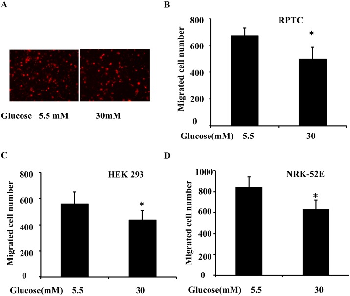
Fig 2. High glucose inhibits transwell migration in cultured kidney tubular cells.
RPTC, NRK-52E and HEK 293 cells were cultured for 2 days in low glucose (5.5 mM) or high glucose (30 mM) medium, and then used for a transwell migration experiment. (A) Representative PI staining of migratory cells was recorded with a fluorescence microscope. A total of 3x10⁵ RPTC were seeded in a transwell insert, which was put in a 24-well plate containing 600 μL low glucose or high glucose medium for 6 h. The cells that migrated to the undersurface were stained with PI and counted. (B) Migratory cells attached to the undersurface were counted after PI staining in RPTC. (C) Migratory cells attached to the undersurface were counted after PI staining in HEK 293 cells. (D) Migratory cells attached to the undersurface were counted after PI staining in NRK-52E. Data are expressed as the mean ± S.D. (n = 4). *, p<0.05 versus the low glucose group.

