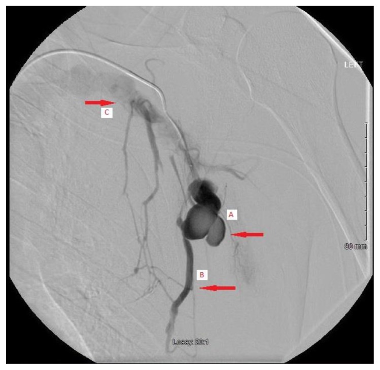
Figure 2.
A 52 year old male status post stabbing with a traumatic left lateral thoracic artery pseudoaneurysm.
Findings: Image: A lobulated pseudoaneurysm of the left lateral thoracic artery (A). Early venous filling his demonstrated (B) indicating arteriovenous fistula formation. Contrast is then seen returning into the central venous system (C).
Technique: Spot radiograph during the venous phase of digital subtraction angiography sequence with injection of iohexol contrast solution through a Renegade Hi-flo microcatheter.

