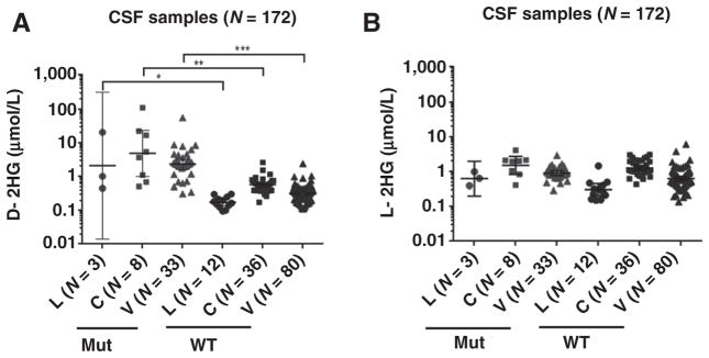
Figure 3.
The levels of D-2HG detected in CSF from lumbar (L), cisternal (C), or ventricular (V) draws were significantly different between WT and mutant IDH patient groups. A, Student t test comparisons show significant differences in the levels of D-2HG found in the CSF of patients with WT or Mut IDH gliomas from lumbar, P = 0.0283, cisternal, P = 0.0022, and ventricular, P = 0.0002, draws. B, There are no significant differences in L-2HG levels between WT and mutant groups. Geometric means with 95% CI are indicated. The number (N) of samples is indicated in the parentheses.

