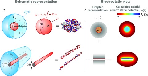
FIG. 1.
(a) Schematic illustration of our model of a charged globular macromolecule (red sphere) and a linear polyelectrolyte (red cylinder) immersed in an electrolyte (blue). Grey spheres denote protons both in free solution and in the interior dielectric environment of the globular molecule. The surface integral of an electric field, E, over the sphere or cylinder yields the true net (regulated) charge of the molecule, . Far away from the molecule, the corresponding surface integral in the electrolyte goes to zero, as a result of electroneutrality within the domain enclosing both molecule and electrolyte. and —volumetric and surface charge densities in the spherical and cylindrical cases, respectively; and —static dielectric constants of the protein interior and external electrolyte, respectively; —Debye length; R—radius of the sphere or cylinder representing the molecule. (b) Left: Graphic representation of a spherical and cylindrical molecule in 2D. The dashed line denotes the axis of cylindrical symmetry and the grey shaded region depicts the counterion density surrounding the molecule. Right: Spatial electrostatic potentials, for a spherical and cylindrical molecule calculated using the non-linear PB equation. The cases presented are those of Gusβ (top) and 60 bp dsDNA (bottom) in 1 mM monovalent salt, pH 8.8.

