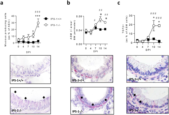
Figure 4.

IPS-1−/− mice exhibit elevated airway remodelling in acute viral infection. (a) Top panel: Number of mucous-producing AECs. Bottom panel: representative micrograph (40x magnification) of Periodic acid–Schiff staining. (b) Top panel: Airway smooth muscle (ASM) area. Bottom panel: representative micrograph (40x magnification) of alpha actin smooth muscle immunostaining. (c) Top panel: Percentage of ASM cells expressing TGFβ. Bottom panel: representative micrograph (40x magnification) of TGFβ immunostaining. Vehicle control mice were sacrificed on day 7 of life (i.e. 0 dpi). Experiments were performed twice with 5 to 7 mice per group. Data are mean and the standard error of the mean. *,**,***,****Denotes significance between WT and IPS-1−/− infected mice. #,##,###Denotes significance between infected and vehicle IPS-1−/− mice.
