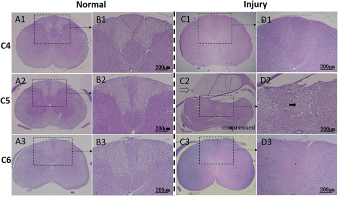
Figure 2.

Histological features of normal and injured cord by hematoxylin erosion (H&E) staining. The normal cord with sham compression showed structural integrity at C5 level (A2) as well as the adjacent C4 (A1) and C6 (A3) level. Butterfly-like gray matter could be clearly identified (A1–3). Distinguished neural fiber tracts derived from dorsal horn (B1–3) could be observed; The compressor (hollow arrow) at C5 level expanded its volume, induced compression injury to the cord, and led to notable structural deformation and disorganization (C2 and D2). Evident tissue loss and diffuse vacuolization (solid arrow) of the posterior funiculus in white matter were verified at the C5 injury level (D2). By comparison, the adjacent levels, C4 (C1, D1) and C6 (C3, D3), with integral and organized structures, which were similar with the normal cord (A1–3 and B1–3), showed no distinguishable pathological changes.
