
Figure 1.
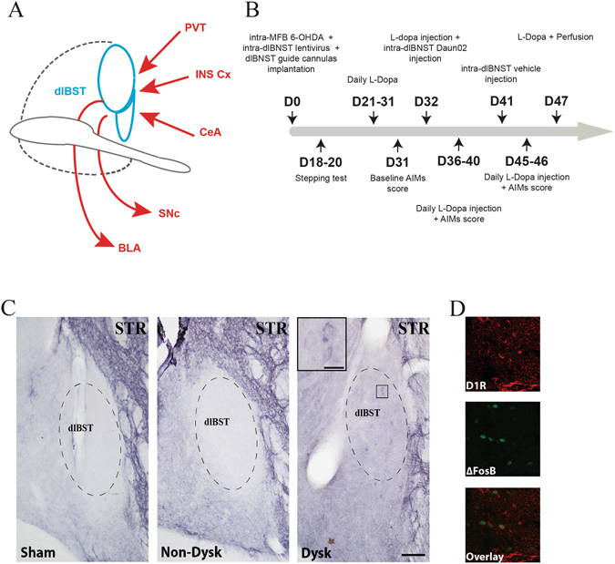
D1R expression in the dlBST. (A) Schematic summary of major known connections of the dlBST. BLA, basolateral nucleus of the amygdala; CeA, central nucleus of the amygdala; INS Cx, insular cortex; PVT, paraventricular nucleus of the thalamus; SNc, substantia nigra pars compacta 24, 43, 44. (B) Timeline of experimental manipulations. D, day. (C) Representative dlBST mapping of D1R expression (dashed lines) in sham-operated (sham), 6-OHDA-lesioned (Non-Dysk) and L-Dopa-treated dyskinetic 6-OHDA-lesioned rats (Dysk) (scale bar: 300 µm) with an inset showing a magnification of D1R expression in dyskinetic condition (scale bar: 5 µm) (STR = Striatum). (D) Representative insets (scale bar: 20 µm) showing D1R, ∆FosB and co-localization of D1R/∆FosB expression.