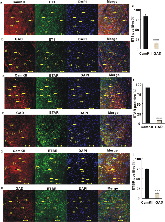
Figure 3.

ET1 and its receptors (ETARs and ETBRs) are mainly present at glutamatergic neurons in the BLA. (a) Coronal sections of the BLA of C57 mice were stained with anti-CamKII antibody, anti-ET1 antibody and DAPI (blue). Most the right image shows combinations of red (CamKII), green (ET1) and blue (DAPI) channels. Unfilled triangles, ET1-positive neurons without CamKII; arrows, ET1- and CamKII-positive neurons. (b) Coronal sections of the BLA were stained with anti-GAD antibody, anti-ET1 antibody and DAPI. Most the right image shows combinations of red (GAD), green (ET1) and blue (DAPI) channels. Unfilled triangles, ET1-positive neurons without GAD; arrows, ET1- and GAD-positive neurons.(c) Quantitative analysis of pyramidal neurons and GABAergic neurons that are positive for ET1. (d) Coronal sections of the BLA were stained with anti-CamKII antibody, anti-ETAR antibody and DAPI. Most the right image shows combinations of red (CamKII), green (ETAR) and blue (DAPI) channels. Unfilled triangles, ETAR-positive neurons without CamKII; arrows, ETAR- and CamKII-positive neurons. (e) Coronal sections of the BLA were stained with anti-GAD antibody, anti-ETAR antibody and DAPI. Most the right image shows combinations of red (GAD), green (ET1) and blue (DAPI) channels. (f) Quantitative analysis of pyramidal neurons and GABAergic neurons that were positive for ETAR. Unfilled triangles, ETAR-positive neurons without GAD; arrows, ETAR- and GAD-positive neurons. (g) Coronal sections of the BLA were stained with anti-CamKII antibody, anti-ETBR antibody and DAPI. Most the right image shows combinations of red (CamKII), green (ETBR) and blue (DAPI) channels. Unfilled triangles, ETBR-positive neurons without CamKII; arrows, ETBR- and CamKII-positive neurons. (h) Coronal sections of the BLA were stained with anti-GAD antibody, anti-ETBR antibody and DAPI. Most the right image shows combinations of red (GAD), green (ETBR) and blue (DAPI) channels. Unfilled triangles, ETBR-positive neurons without GAD; arrows, ETBR- and GAD-positive neurons. (i) Quantitative analysis of pyramidal neurons and GABAergic neurons that were positive for ETBR. Scale Bar = 50 µm. Shown are means ± SE; n = 40 for CamKII-positive cells of 10 independent sections; n = 10 for GAD-positive cells of 10 independent sections. The 10 independent sections of each group were randomly selected from 5 mice. ***P < 0.001, Student’s t test for 3c, 3f, 3i.
