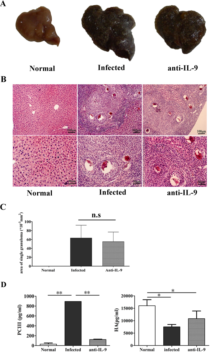
Figure 5.

Roles of anti-IL-9 in S. japonicum infection-induced hepatic granulomatous and fibrosing inflammation. Female C57BL/6 mice were randomly divided into three groups: normal group, infected group and anti-IL-9 group. The infected group and anti-IL-9 group were infected with 40 ± 5 S. japonicum cercariae per mouse. From week 5 post-infection, the control IgG mAb or anti-IL-9 mAb were injected intraperitoneally into the infected group and anti-IL-9 group mice every 3 days, for a total of 6 times, respectively. These mice were sacrificed at 8 weeks post-infection. Mice livers were fixed in paraformaldehyde, embedded in paraffin, and then sliced and examined by haematoxylin and eosin staining. (A) The gross appearance of livers from the three groups. (B) Representative photomicrographs are shown at x100 magnification for upper panels and ×200 for lower panels. (C) Sizes of single egg granuloma were measured by computer-assisted morphometric analysis. Only granulomas with a visible central egg were analysed for accuracy. The results are expressed as mean ± SD (6 mice per group); n.s, P > 0.05. (D) Levels of pro-collagen type III (PC-III, left) and hyaluronic acid (HA, right) in the serum of mice were detected by ELISA. The results are expressed as mean ± SD; *P < 0.05, **P < 0.01. One representative result of 3 to 5 independent results is shown.
