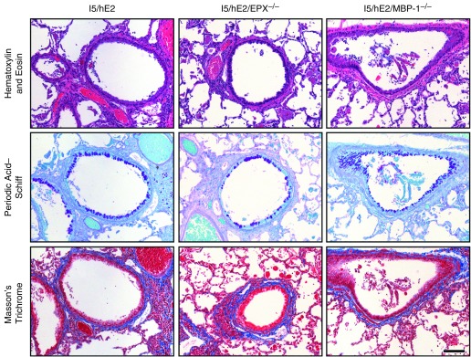
Figure 6.
The loss of either of the abundant secondary granule proteins (i.e., eosinophil peroxidase [EPX] or major basic protein-1 [MBP-1]) has nominal but differential effects on the induced histopathologies/remodeling occurring in I5/hE2 mice. Parasagittal lung sections of transgenic/compound granule protein gene knockout mice were stained to evaluate the dependency of the pulmonary histopathologies occurring in the parental I5/hE2 model on EPX (i.e., I5/hE2/EPX−/−) and MBP-1 (i.e., I5/hE2/MBP-1−/−). Hematoxylin and eosin and Masson’s trichrome staining demonstrated that the loss of either granule protein had little to no effect on the leukocyte tissue infiltration or the induced lung structural changes (including pulmonary fibrosis [blue-staining extracellular matrix regions in Masson’s trichrome–stained lung sections]) occurring in the parental I5/hE2 model. In contrast, periodic acid–Schiff staining of lung sections for goblet cell metaplasia/epithelial cell mucin accumulation (dark-purple-staining cells) demonstrated a differential effect regarding the loss of these granule proteins with the loss of EPX and not MBP-1, resulting in a greater than 50% reduction in goblet cell metaplasia/epithelial cell mucin accumulation relative to the levels observed in I5/hE2 mice. Scale bar = 100 μm. The cohort size of each group (i.e., I5/hE2, I5/hE2/EPX−/−, and I5/hE2/MBP-1−/−) was n = 3–5 mice.

