
Figure 7.
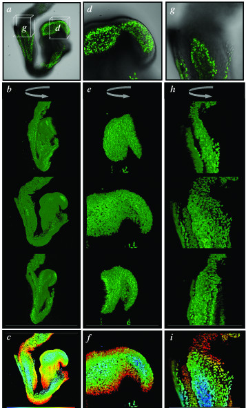
High-resolution 3-dimensional imaging of fixed CAG::H2B-EGFP transgenic embryos. Confocal images of an E8.5 CAG::H2B-EGFP transgenic embryo fixed in 4% paraformaldehyde for 72 hours, then washed, stored and imaged in PBS. Low-magnification views and reconstructions of whole embryo (a-c). Boxes in a designate region imaged in d and g. High-magnification views of the headfolds (d-f) and posterior primitive streak and proximal allantois (g-i). Single xy images (a, d and g) from the z-stacks used to computationally render the data sets. These images are overlayed onto the bright field channel so as to display the outline of the embryo. Rotations through the rendered z-stacks displayed at 45° intervals (b, e and h). Color-coded depth projections of each of the z-stacks (c, f and i).