
Fig. 3.
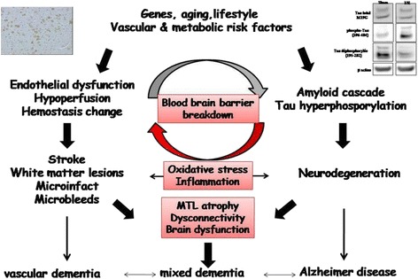
Pathophysiological mechanisms of interactions between vascular and degenerative processes in both vascular and mixed dementia (Adapted from [40]). MTL medial temporal lobe. The figure integrates an image of western blot (tau expression after middle cerebral artery occlusion in mice) and immunohistochemistry (amyloid peptide expression after middle cerebral artery occlusion in rat)