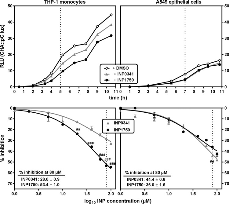
FIG 3.
Influence of INP0341 and INP1750 on iT3SS transcription. (Top panels) iT3SS transcriptional activation of the exsCEBA operon, followed by measuring the bioluminescence signal of the reporter strain CHA pC::lux over time, under control conditions (0.8% DMSO) or in the presence of 80 μM INP0341 or INP1750. Bacteria (10/cell) were cultured in the presence of THP-1 monocytes in RPMI 1640 medium (left) or of A549 epithelial cells in DMEM (right). Bioluminescence is expressed in arbitrary units (RLU), which correspond to 10−4-fold the actual readings. The dotted lines indicate the time selected in the bottom panels. (Bottom panels) Inhibition of the transcriptional activation of exsCEBA operon by increasing concentrations of INP0341 or INP1750, as measured after 5 h of incubation of CHA pC::lux (10 bacteria/cell) in the presence of THP-1 monocytes (left) or after 7 h of incubation in the presence of A549 cells (right). Values are expressed in percentage of inhibition of the bioluminescence signal recorded in the absence of inhibitors. All values are means ± the SEM of two or three experiments. Statistical analyses were performed (two-way ANOVA, Bonferroni posttest; comparison between cells incubated with INP0341 and cells incubated with INP1750 [###, P < 0.001; ##, P < 0.01]).

