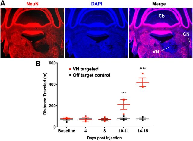
Figure 6.
Hyperactivity is induced by lesioning the vestibular nucleus. A, Coronal section showing a unilateral AAV-Cre-induced lesion (loss of NeuN-labeled neurons, red) in the VN of R26DTA/DTA mice. Hoechst-33342 counterstain (blue). CN, Cochlear nucleus; Cb, cerebellum. B, Correctly targeted VN-lesioned mice become hyperactive. Controls are lesions from shallower injections of virus into the ventral cerebellum immediately adjacent and dorsal to the VN. Data are mean ± SEM (n = 3 VN targeted mice, n = 5 controls). ***p = 0.0003, two-way repeated-measures ANOVA with Tukey multiple-comparisons test. ****p < 0.0001, two-way repeated-measures ANOVA with Tukey multiple-comparisons test.

