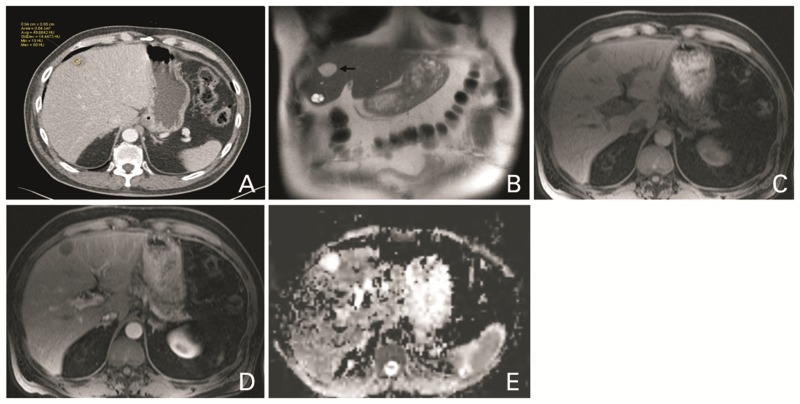
Figure 3.
A 68-year-old man with ciliated hepatic foregut cyst. Contrast-enhanced CT (A) shows a hypodense lesion in segment 4B, with attenuation slightly more than water (49 Hounsfield Unit). The lesion (arrow) is bright on T2 WI (B) but not as bright as a cyst also seen in segment 4B. The lesion is hypointense on T1 weighted image (C) with no enhancement after contrast injection (D). There is no associated diffusion restriction, as can be seen on the ADC image (E).

