
Fig. 3.
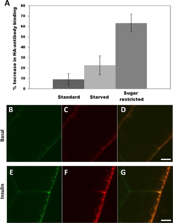
Drosophila harbor the machinery to mediate an insulin-dependent sugar uptake response. (A) Surface exposure of HA-GLUT4 upon insulin stimulation. Fat bodies were collected from larvae reared on different dietary regimes, as indicated. After immunostaining of nonpermeabilized fat body cells, values were determined by measuring Alexa 647 conjugated anti-HA antibody fluorescence and averaging calculations of corrected integrated density. Values are expressed as percent of HA-GLUT4 fluorescence of insulin stimulation over basal conditions. Fat bodies were collected from three animals for each dietary regime. (B–G) Confocal microscopy of HA-GLUT4-GFP expression in fat body cells from animals reared on a sugar-restricted diet in the absence or presence of insulin. (B, C, and D) Basal conditions; (E, F, and G) after addition of 0.1 U/mL insulin. GLUT4 was visualized in nonpermeabilized cells by GFP fluorescence (green B and E) or with anti-HA antibody (red C and F) to monitor membrane translocation. Scale bar is 5 μm. Note increase in GLUT4 at the cell surface (red F). From Crivat, G., Lizunov, V. A, Li, C. R., Stenkula, K. G., Zimmerberg, J., Cushman, S. W., & Pick, L. (2013). Insulin stimulates translocation of human GLUT4 to the membrane in fat bodies of transgenic Drosophila melanogaster. PloS One, 8(11), e77953. doi:10.1371/journal.pone.0077953.