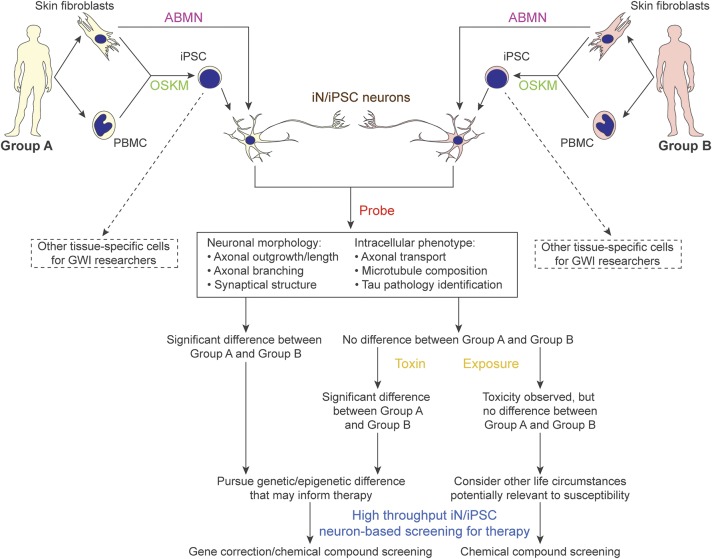
Figure 3. Schematic flow chart for planned experimental regimen.
Group A, Gulf War (GW) veterans without GW illness (GWI); group B, GW veterans with GWI. The types of assessments that will be done on the cells depend on the hypothesis being pursued. Our initial plan, to probe phenotypes mentioned in the chart, will be to measure lengths of axons after toxicant exposures, as well as to count branches along the axons as normalized per 100 μm, quantify synapses by performing patch-clamp recording to quantify spontaneous activities and by immunostaining for synaptophysin or synapsin, examine axonal transport by performing live-cell imaging using fluorescent markers for various organelles, and examine microtubule structure by immunocytochemistry and Western blotting using antibodies to various tubulin variants and microtubule-associated proteins. We are especially interested in studying tau, a microtubule-associated protein that dissociates from microtubules to form abnormal filaments in a number of human neurodegenerative diseases, a pathologic phenomenon not well reflected in rodent neurons.36 One-way analyses of variance will be used for all the statistic quantitations with n > 50. ABMN = transcription factors Ascl1, Brn2, Myt1l, and NeuroD1; fibs = fibroblasts; iN = induced neurons; iPSC = induced pluripotent stem cells; OSKM = transcription factors Oct4, Sox2, Klf4, and c-Myc; PBMC = peripheral blood mononuclear cells.

