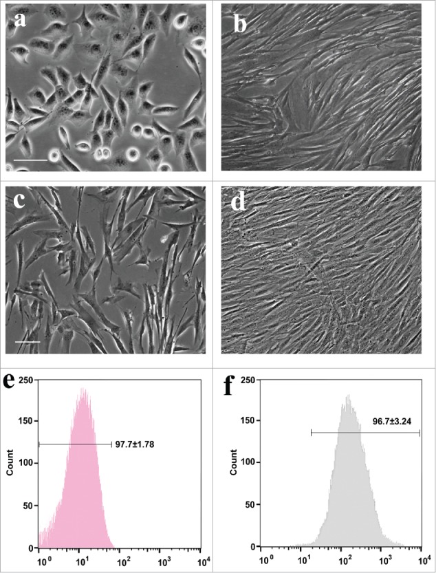
Figure 2.

Phase-contrast images and flow cytometric ADSCs. (A, B) The morphology of primary ADSCs at 3 and 7 d in vitro, respectively. (C, D) Purified ADSCs at 2 and 5 d in vitro. (E, F) Rat ADSCs at 2 passages were harvested for flow cytometric analysis with CD31 and CD44.
