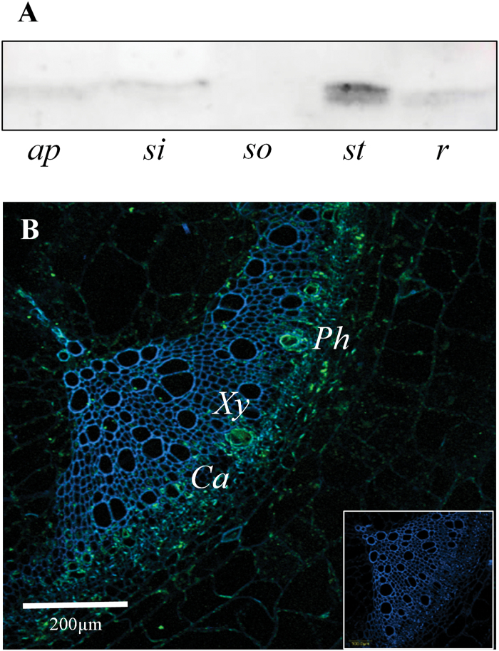
Fig. 1.
Spatial distribution of Cyp1 proteins in different organs. (A) Anti-cyclophilin western-blot analyses on protein extracts from different tomato organs using an anti-AtCyp18-3/ROC1 antiserum. Proteins were extracted from the following organs: shoot apex (ap), sink leaf (si), source leaf (so), stem (st), and root (r). (B) SlCyp1 immunolocalization in a transverse section of tomato stem. The green signal indicates SlCyp1 and the blue signal indicates xylem autofluorescence. Note that SlCyp1 localizes mainly to the phloem (Ph), cambium (Ca), and developing xylem (Xy) vessels. Inset: negative control. Images were obtained using an Olympus IX-81 confocal microscope.

