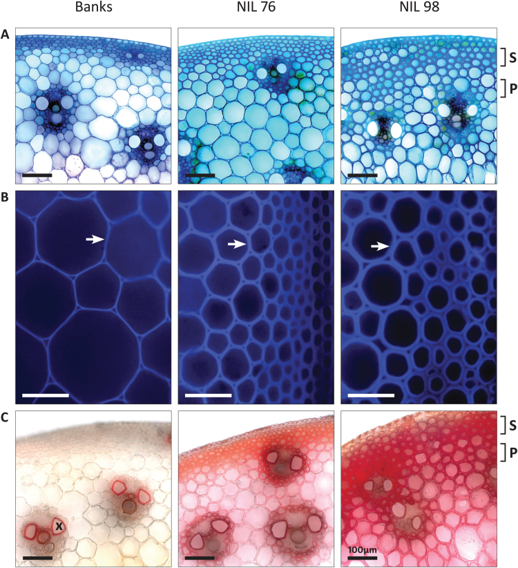
Fig. 3.
Microscopic analysis of stem cross-sections of cultivar Banks, NIL76, and NIL98 from the midpoint of internode P-2 at late stem elongation. (A) Toluidine Blue stain with bright-field illumination (20×). (B) Unstained, autofluorescence (20×). (C) Mäule stain with bright-field illumination (20×). Sclerenchyma (S) and parenchyma (P) cell layers, and xylem vessel (X) are indicated. Cell wall thickening is indicated by the arrows. Scale bars=100 µm.

