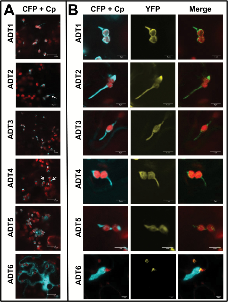
Fig. 2.
Subcellular localization of ADT–FP fusion proteins and co-localization with TP-ssRuBisCO–YFP. (A) ADT–CFP subcellular localization patterns. ADT1–ADT5 localized to stroma and to areas seemingly close to the chloroplast just outside of the autofluorescence signal generated by chlorophyll. They often appear either in thread-like structures (e.g. the arrow in ADT2) or globular structures (e.g. the arrows in ADT4). The ADT6–CFP pattern is distinctly different, showing a cytosolic distribution. Images were taken at a lower magnification to allow observation of the CFP signal relative to several chloroplasts. (B) Close-ups of ADT–CFP subcellular localization patterns in relation to TP-ssRuBisCO–YFP. In contrast to the chlorophyll autofluorescence, the TP-ssRuBisCO–YFP is a stroma-specific marker that visualizes all stroma-filled areas within the chloroplast including stromules. ADT1–ADT5 are found within the main body of chloroplast and in stromules, while ADT6 is found within the cytosol and does not co-localize with TP-ssRuBisCO–YFP.

