Fig. 6.
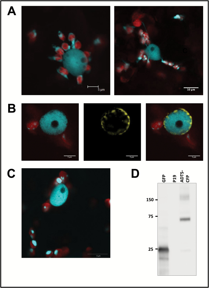
ADT5 is found in the nucleus. ADT5–CFP proteins are unique as they are the only full-length ADT proteins that were found in the nucleus. (A) Nuclei show a close association with chloroplasts (left) or with stromules of chloroplasts (right). Both images show ADT5–CFP within nuclei. (B) Co-localization of ADT5–CFP with NUP1–YFP. To determine if ADT5–CFP localizes to the nucleus, it was co-expressed with NUP1–YFP in N. benthamiana. Images of chlorophyll fluorescence and ADT5–CFP are shown merged (left). NUP1–YFP is shown alone (middle) and merged with ADT5–CFP and chlorophyll fluorescence (right). NUP1–YFP localizes to the nuclear membrane and surrounds ADT5–CFP, confirming that it localizes to the nucleus. (C) ADT5–CFP transiently expressed with its native ADT5 promoter also localizes to the nucleus. (D) Western blot of ADT5–CFP (calculated size 73.9 kDa) expressed with its native promoter and visualized with a GFP antibody is detected at its expected size. As negative controls, proteins isolated from leaves transformed with GFP (25 kDa) and p19 are shown. Total soluble protein was isolated from transiently transformed leaves, and 10 μg of total soluble protein was size separated by 10% SDS–PAGE. Sizes of the protein ladder are given in kDa.

