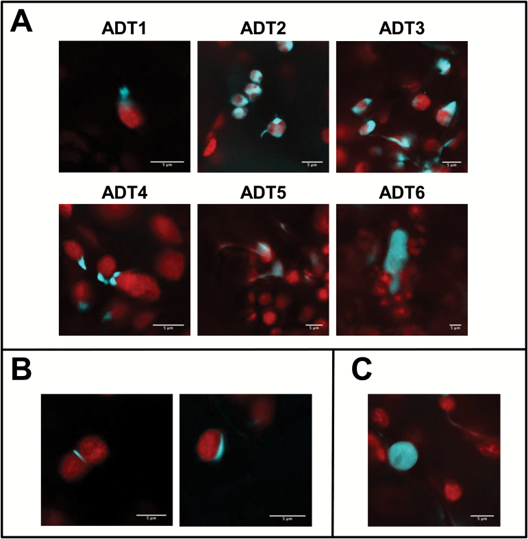
Fig. 8.
ADT localization to the stroma and stromules, the chloroplast equatorial plane, and the nucleus can also be detected in A. thaliana. To test if the ADT patterns determined in N. benthamiana reflect expression in A. thaliana, all six ADT–CFP fusion proteins were transiently expressed in A. thaliana Col-0. All images show a merge of chlorophyll and CFP fluorescence. (A) ADT1–CFP through ADT5–CFP localize to stroma and structures resembling stromules of varying shapes and lengths, with varying levels of fluorescence in the stroma. ADT6–CFP localizes outside of chloroplasts in the cytosol. (B) Chloroplast division patterns for ADT2–CFP. (C) Nuclear localization of ADT5–CFP.

