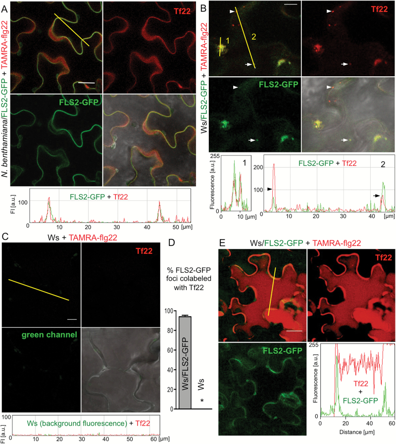
Fig. 4.
flg22 is internalized together with FLS2. Leaf discs of N. benthamiana (A) and Arabidopsis Ws expressing FLS2–GFP (B, E) or control Ws (C) were floated on 2 μM TAMRA–flg22 (Tf22) for the indicated time, washed in water and imaged by confocal microscopy. Images in red (TAMRA) and green (GFP) channels, and their composite with or without DIC image are shown. Graphs show fluorescence profiles along yellow line. Bar: 20 μm in (A, E) and 10 μm in (B, C). These experiments were repeated three to four times. (A) TAMRA–flg22 colocalizes at plasma membrane with FLS2–GFP after several minutes to 1 h incubation. See additional images in Supplementary Figs S5 and S6. (B) TAMRA–flg22 is internalized and colocalizes with FLS2–GFP in vesicles (line 2, two vesicles along the line marked with an arrowhead and an arrow) and MVBs (line 1) after 1–2 h incubation. See additional images in Supplementary Fig. S7. (C) TAMRA–flg22 does not enter wild-type Ws cells that lack FLS2 after 1–2 h incubation. See additional images in Supplementary Figs S6 and S7. (D) TAMRA–flg22 colocalizes with FLS2–GFP after floating Ws/FLS2–GFP leaf discs on peptide solution for 1–2 h. Fluorescent foci were counted in 16 picture areas (20 μm × 20 μm) for each genotype. Total number of foci: Ws/FLS2–GFP: 152 green (FLS2–GFP), 161 red (Tf22), 143 overlap; Ws: 2 green, 5 red, 0 overlap. Error bars, SE, *P<0.0001 (t-test, n=16). (E) After overnight incubation, TAMRA–flg22 fills the cells, whereas FLS2–GFP is mostly detected at the plasma membrane.

