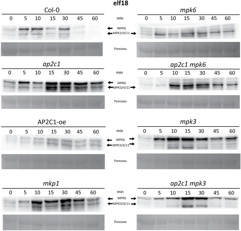
Fig. 2.
AP2C1 controls elf18-induced MAPK activation. Western blotting with p44/42 antibodies after application of 1 µM elf18 on seedlings. Profiling of MAPK activation by an immunological assay that detects phosphorylation of the MAPKs. MPK6 and MPK3/4/11 corresponding immunoreactive bands are indicated by arrows in the top panels. Ponceau staining was used to estimate equal loading (bottom panels).

