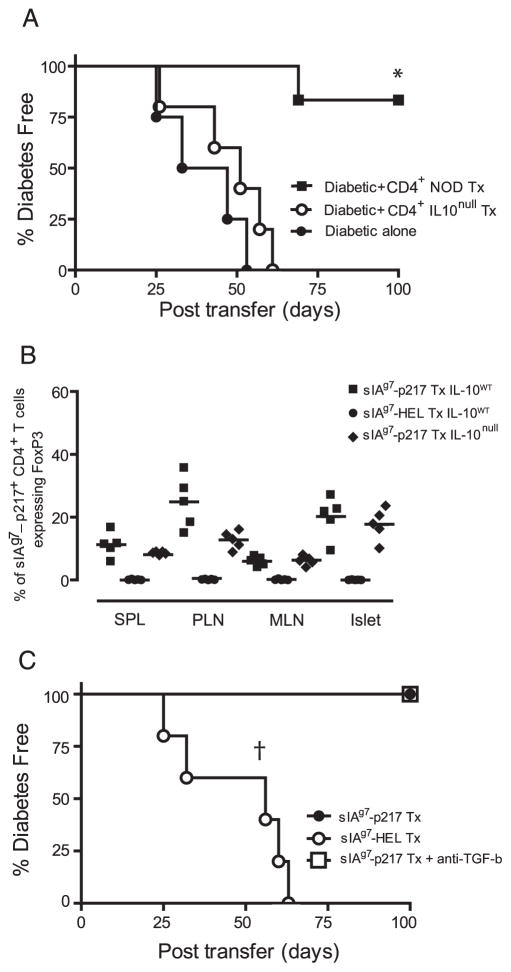
FIGURE 6.
Protection mediated by sIAg7-GADp217 treatment is IL-10-dependent. A, Groups of NOD.scid mice (n = 4) received diabetogenic splenocytes alone or a mixture of purified splenic CD4+ T cells isolated from NOD (n = 6) or NOD.IL-10null (n = 5) female mice treated with sIAg7-GADp217, and diabetes was monitored. *, p = 0.001, by Kaplan-Meier log-rank test for CD4+ T cells from sIAg7-GADp217 treated (Tx) NOD vs NOD.IL10null mice or diabetogenic splenocytes alone. B, Frequency ± SD of sIAg7-GADp217 multimer-staining CD4+ T cells ex vivo expressing FoxP3 in the spleen, PLN, MLN, and islets of n = 5 individual NOD (IL-10wt) or NOD.IL-10null (IL-10null) female mice treated with sIAg7-GADp217 or sIAg7-HEL. C, CD4+ T cells (5 × 10⁶) isolated from the PLN of NOD female mice vaccinated at 12 wk of age with sIAg7-GADp217 or sIAg7-HEL were mixed with splenocytes from diabetic NOD donors (5 × 10⁶) and transferred into groups of n = 5 NOD.scid mice. One group of NOD.scid recipients of CD4+ T cells isolated from sIAg7-GADp217 vaccinated animals also received a TGF-β-neutralizing Ab. †, p = 0.0002, by Kaplan-Meier log-rank test, for recipients of CD4+ T cells from sIAg7-HEL vaccinated mice vs recipients of CD4+ T cells from sIAg7-GADp217 vaccinated mice with or without anti-TGF-β Ab.

