Figure 3.

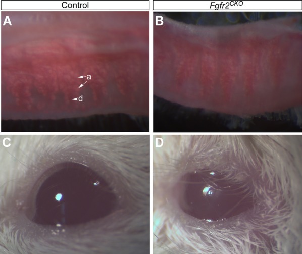
MG atrophy and signs of DED in Fgfr2CKO mice. (A, B) ORO lipid staining in control and Fgfr2CKO mice. Upper eyelids were dissected from control mice (fed regular chow) and Fgfr2CKO mice (fed Dox chow for 10 days) and subjected for whole-mount ORO staining to view lipid (meibum) production by MG acini (arrows labeled a). Meibum secreted into main duct (arrowhead labeled d) was stained in a dark-red color in control mice (A). Compared with control mice, Fgfr2CKO mice exhibited a reduction of MG acinar area and meibum production (B). (C, D) Compared with control mice (C), Fgfr2CKO mice showed signs of ocular irritation, including loss of corneal luster and macerated eyelids, after extended (2 to 3 weeks) Dox chow feeding (D). Such signs of ocular irritation were likely due to increased tear evaporation and tear film instability.
