Figure 7.
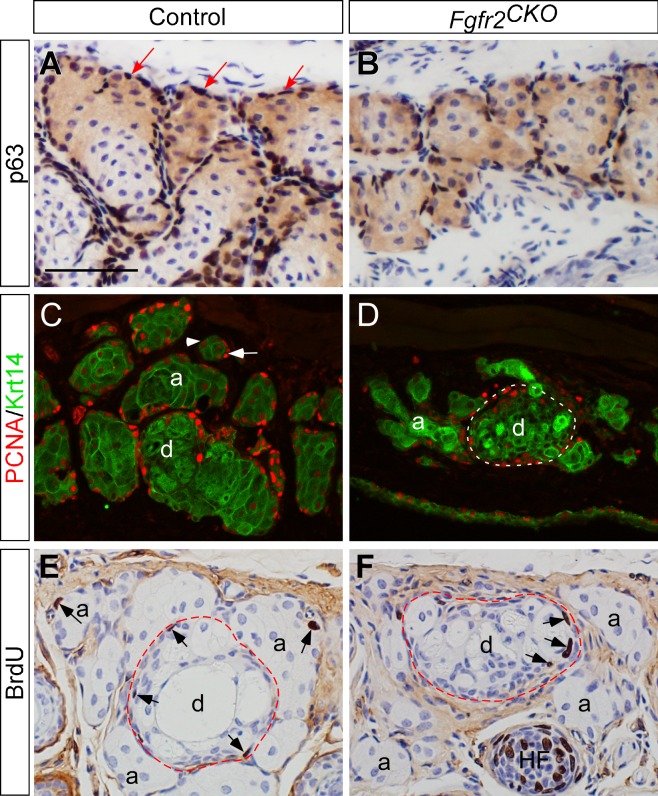
Expression of basal and proliferating cell markers. (A, B) Transcription factor p63 in MGs detected by immunohistochemistry on crysections. The basal cells in MG acini (dark brown nuclei indicated by arrows in A) and ducts (data not shown) express p63 (A). In FGFR2CKO mice fed with Dox for 4 days, the number of basal cells around the MG acini was markedly reduced (B). (C, D) Immunofluorescence of Krt14 (green) and proliferative cell nuclear antigen (PCNA) (red). Krt14 expression marks the areas of MGs in eyelids. In control mice (C), intense PCNA immunofluorescence was found in acinar (a) and ductal (d) basal epithelial cell nuclei (red as indicated by white arrow in C), suggesting that these cells are in the proliferative state. Within a MG acinus, PCNA immune intensity decreases when basal cells are differentiated into meibocytes (arrowhead in C). In Fgfr2CKO mice induced by Dox for 1 week, PCNA-positive nuclei were rarely seen in acinar basal cells, but detected in ductal basal layer (white broken-line circle in D). (E, F) BrdU-labeled cells were found in MG acinar and ductal basal cell layer in control mice (arrows in E). In Fgfg2CKO mice fed with Dox for 6 days, BrdU incorporation was seen in MG ductal basal cells (arrows in F), but rarely detected in the acini. HF, hair follicle.

