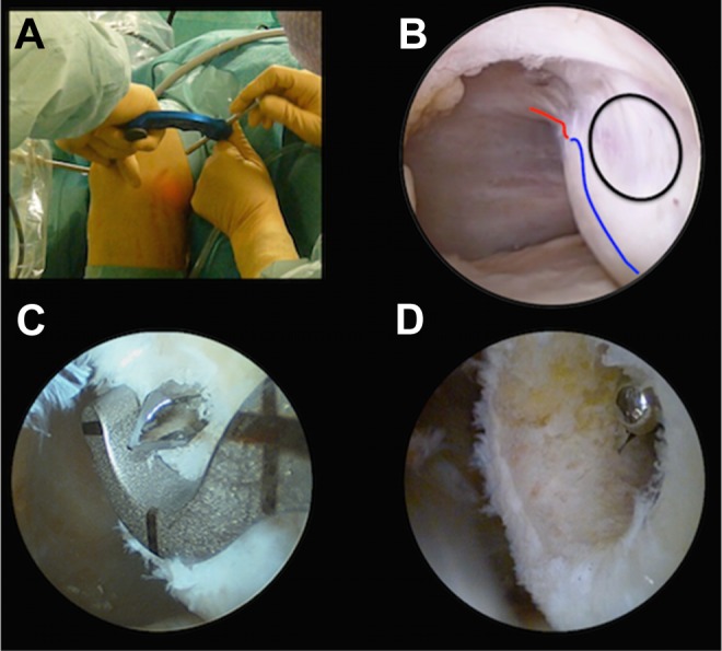
Figure 2.

Left knee arthroscopy with anteromedial (AM) viewing. (A) External view showing positioning of specific outside-in femoral guide through the anterolateral portal. (B) Arthroscopic landmark with the camera through the AM portal: black line, ring target of the guide; red line, notch outlet; blue line, anterior-inferior cartilaginous limit. (C) Femoral anterior cruciate ligament footprint guide and emerging drill pin. (D) Femoral tunnel.
