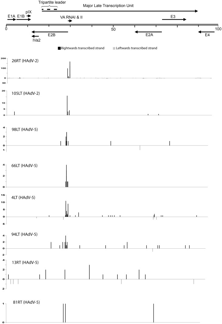
Fig 7. Distribution of small RNAs reads from the HAdV genome in patient tonsillar lymphocytes.
A schematic drawing showing the position of HAdV transcription units is shown at the top. Reads derived from the rightwards-transcribed strand is shown with black boxes and reads derived from the leftward-transcribed strand is shown as grey boxes. The number of reads is shown on the y-axis. The abbreviation of patient samples was as follow: first patient number (Table 1) followed by right (R) or left (L) tonsil followed by the origin of tonsillar cells (T or B lymphocytes). In the patient samples diagnosed with a HAdV-3 infection the virus-specific small RNA accumulation was at a background level (data not shown).

