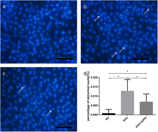
Fig 2. The morphologic changes in NRK-52E cells were displayed by Hoechst 33258 staining.
NRK-52E cells were treated with high glucose with or without adiponectin for indicated time. Then, fluorescence images were taken after Hoechst 33258 staining. Fragmented and pycnotic nuclei were emphasized by white arrows. (200×) (a) control group. (b) SHG group. (c) SHG+ADPN group. (d) Histogram represents the percentage of apoptotic cells. *P<0.01.

