
Figure 1.
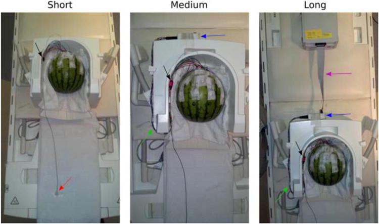
Watermelon phantom with 1 ECG electrode (red arrow) and 21 scalp EEG electrodes placed in accordance with the International 10-20 system, for Experiment 1. The short, medium and long wire configurations described in the Methods are shown in the left, center and right panels, respectively. The black arrow indicates the black Molex connectors, which connect to the braided wires indicated with the green arrow. The blue arrow indicates the Brain Products interface box, which connects to the ribbon cable (purple arrow) that leads to the amplifier. A short ribbon cable is shown here in order to fit the amplifier into the photograph frame, whereas a long ribbon cable was used in the actual experiment.