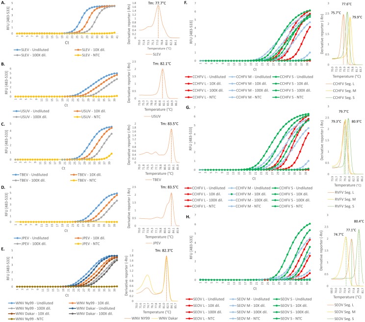
Fig 2. Real-time PCR assays of members from the Flaviviridae and Bunyaviridae families.
Amplification and melting curves for five different flaviviruses species are shown. Each sample was tested undiluted, with a 10-fold dilution and with a 100-fold dilution. (A) St. Louis encephalitis virus (SLEV). (B) Usutu virus (USUV). (C) Tick-borne encephalitis virus (TBEV). (D) Japanese encephalitis virus (JPEV). (E) West Nile virus (WNV; 2 strains, NY99 and Dakar). The right half of the panel shows the amplification and melting curves of the different genomic segments of the members from the Bunyaviridae family tested in this study. (F) Crimean-Congo hemorrhagic fever virus (CCHFV). (G) Rift Valley fever virus (RVFV). (H) Seoul virus (SEOV). NTC, no template control; RFU, relative fluorescence units; Ct, cycle threshold; Dil., dilution; Seg., Segment.

